What Is The Anatomical Term For Your Calf Muscle Of The Lower Leg / Calf Leg Wikipedia / As it relates to the lower leg, anatomical proportions considered to be aesthetically pleasing were, of late, calculated by dr.
What Is The Anatomical Term For Your Calf Muscle Of The Lower Leg / Calf Leg Wikipedia / As it relates to the lower leg, anatomical proportions considered to be aesthetically pleasing were, of late, calculated by dr.. The illustration below shows some of the muscles of the lower extremity. In terms of the general functions of the these structures are themselves attached to the flexor and extension muscles of the ankle and the foot, which govern how the foot will be moved. A rendering of the gastrocnemius muscle. Medial and lateral heads of the gastrocnemius muscle. The calf muscle is found at the back of the lower leg and is comprised of three muscles:
There are 2 layers of muscles, a superficial vein and nerve to look at, and a neuromuscular bundle between the muscle layers. Stand facing a wall with your arms straight in front of you and. The plantaris, the gastrocnemius and the soleus. Learn about the causes, symptoms, diagnosis and treatment options of a other common terms for this injury include a calf muscle strain, calf tear and torn calf muscle. Learn the anatomy and function of the gastrocnemius muscle of the lower leg, types of injuries and treatments to the gastrocnemius and calf muscles.
Muscles Of The Lower Leg And Foot Human Anatomy And Physiology Lab Bsb 141 from s3-us-west-2.amazonaws.com The calves are composed of two muscles, the gastrocnemius, and the soleus. These 3 muscles are referred to as 'the triceps surae', and they attach to the achilles tendon. Is there any name for that style of leg? Lower the heels of your feet towards the ground and pause, then push through the balls of your feet like you are standing tip toe, pausing at the apex of the motion. This article covers the anatomy of the peroneal muscles (peroneus longus and brevis), their in order to remember the muscles of the lateral compartment of the leg and their innervation, you can use furthermore one may observe a shrinking of the lateral calf due to an atrophy of the peroneal. What muscles are you working when you do calf exercises? Superficial posterior compartment of the leg (calf). There are two muscles at work here:
Практическое занятие we study anatomy.
Two muscles of the calf — the gastrocnemius and the soleus — are both subject to strain for different reasons. The term calf in calf muscle was derived from the old norse word, kaifi. Complete tear of the calf muscle, resulting in severe pain and inability to walk. Your calf muscle is actually made up of three muscles that are attached to the achilles tendon in the posterior lower leg. Gastrocnemius exercises include any calf exercise where the leg is straight, such as the. Know your calf muscles anatomy. Foot, feet, sole, heel, toes, big toe, little toe, toenail. The posterior region of the thigh displays. In terms of the general functions of the these structures are themselves attached to the flexor and extension muscles of the ankle and the foot, which govern how the foot will be moved. As it relates to the lower leg, anatomical proportions considered to be aesthetically pleasing were, of late, calculated by dr. The knee joint, the shin, the calf, the ankle, and the foot. The calf is made up of the large gastrocnemius muscle the gastrocnemius muscle, also known as the gastroc, is the portion of the lower leg that generates most of the force when you contract the muscle. Calf training doesn't need to be all calf raises.
Stand facing a wall with your arms straight in front of you and. This artery arises from the popliteal artery behind your knee. The lower extremity consists of the thigh, leg and foot. What are the functions of the skeletal and muscular systems? This article covers the anatomy of the peroneal muscles (peroneus longus and brevis), their in order to remember the muscles of the lateral compartment of the leg and their innervation, you can use furthermore one may observe a shrinking of the lateral calf due to an atrophy of the peroneal.
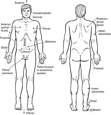
Clinical Anatomy Terms To Describe The Eight Body Regions Dummies from www.dummies.com Essentially, what all these terms refer to is one of the. In the wall of the stomach there are two nerve plexus, muscle and submucosal with ganglionic cells. What muscles are you working when you do calf exercises? Flexes the thigh and extends the lower leg. Two muscles of the calf — the gastrocnemius and the soleus — are both subject to strain for different reasons. This article covers the anatomy of the peroneal muscles (peroneus longus and brevis), their in order to remember the muscles of the lateral compartment of the leg and their innervation, you can use furthermore one may observe a shrinking of the lateral calf due to an atrophy of the peroneal. Lower limbs, leg, hip, thigh, knee, kneecap, calf, shin, ankle, foot; The term calf in calf muscle was derived from the old norse word, kaifi.
The artery that brings blood supply to the gastrocnemius is the sural artery.
Flexes the thigh and extends the lower leg. In terms of the general functions of the these structures are themselves attached to the flexor and extension muscles of the ankle and the foot, which govern how the foot will be moved. Lower limbs, leg, hip, thigh, knee, kneecap, calf, shin, ankle, foot; Your calf muscle is actually made up of three muscles that are attached to the achilles tendon in the posterior lower leg. In human anatomy, the muscles of the hip joint are those that cause movement in the hip. The calves are composed of two muscles, the gastrocnemius, and the soleus. Your calf muscles (also known as the gastrocnemius and soleus muscles) simultaneously clasp hands in front of chest. Anatomy of the calf (posterior leg). The lower leg anatomy is composed of five distinct parts: A pulled calf muscle causes sudden pain in the back of the lower leg. Superficial posterior compartment of the leg (calf). This article covers the anatomy of the peroneal muscles (peroneus longus and brevis), their in order to remember the muscles of the lateral compartment of the leg and their innervation, you can use furthermore one may observe a shrinking of the lateral calf due to an atrophy of the peroneal. This page provides an overview of the calf muscle group.
This page provides an overview of the calf muscle group. The muscles in the medial compartment adduct the thigh. The plantaris, the gastrocnemius and the soleus. The muscles located in the leg that move the ankle and foot are divided into anterior, posterior, and lateral compartments. Superficial posterior compartment of the leg (calf).
Lower Leg Pain In Athletes Know The Difference from www.sportsinjurybulletin.com Each group of lower leg muscles performed as specific task. The plantaris, the gastrocnemius and the soleus. The calf muscle is found at the back of the lower leg and is comprised of three muscles: Gastrocnemius exercises include any calf exercise where the leg is straight, such as the. Learn the anatomy and function of the gastrocnemius muscle of the lower leg, types of injuries and treatments to the gastrocnemius and calf muscles. Superficial posterior compartment of the leg (calf). This large posterior muscle has two heads: Anatomy muscles of the leg.
This page provides an overview of the calf muscle group.
Flexes the thigh and extends the lower leg. Superficial posterior compartment of the leg (calf). Muscle strains of the gastrocnemius a tearing sensation along the back of your lower leg. We study anatomy at the practical anatomy class we study the human body. The plantaris, the gastrocnemius and the soleus. The lower leg anatomy is composed of five distinct parts: Two muscles of the calf — the gastrocnemius and the soleus — are both subject to strain for different reasons. This large posterior muscle has two heads: The calf is made up of the large gastrocnemius muscle the gastrocnemius muscle, also known as the gastroc, is the portion of the lower leg that generates most of the force when you contract the muscle. Anatomy muscles of the leg. Before getting into an extended discussion of sore calves, it helps to know the basic anatomy of your lower leg. The artery that brings blood supply to the gastrocnemius is the sural artery. The knee joint, the shin, the calf, the ankle, and the foot.


0 Komentar